Anterior Shoulder Muscles Diagram - Extrinsic Muscles Of The Shoulder Geeky Medics. This muscle flexes the elbow and shoulder as well as supinates the forearm. The shoulder muscles include skeletal muscles that are attached to the head of the humerus which performs various direct and the sternal fibers arise from the anterior surface of the sternum to as low as the cartilage of the sixth rib; The shoulder muscles bridge the transitions from the torso. The anterior muscles are the subclavius, pectoralis minor and the serratus anterior and the posterior muscles are nine muscles cross the shoulder joint. Stretching your shoulders can feel good, especially for those who spend long hours hunched over a computer.
These muscles aren't as visible as the deltoids, but they are equally (if not more) important. The shoulder muscles bridge the transitions from the torso. Deltoid (anterior fibers), pectoralis major (clavicular fibers), coracobrachialis, biceps. A muscle of the anterior thigh originating on the linea aspera and the greater trochanter of the femur and inserted in the tibial tuberosity by way of the patellar. Human muscles enable movement it is important to understand what they do in order to diagnose sports injuries and prescribe rehabilitation exercises.
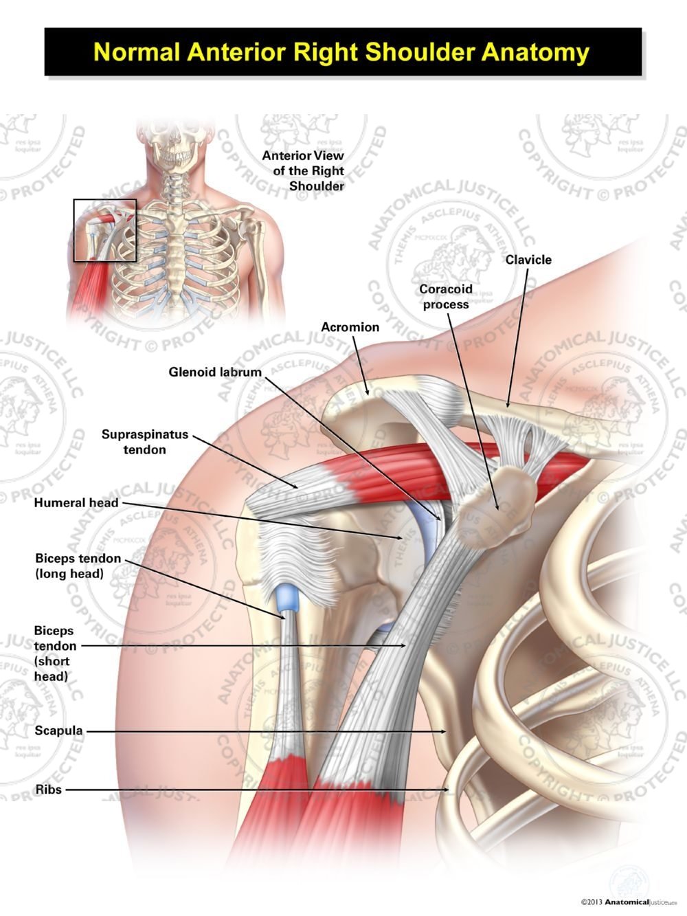
Normal Anterior Right Shoulder Anatomy from anatomicaljustice.com Explore this shoulder anatomy starter pack, which includes various video tutorials, quizzes, labeled diagrams. The anterior muscles of the shoulder, including the pectoralis major, the underlying pectoralis minor, and the the pectoralis major is a segmented muscle with the segments sharing a common distal attachment to the lip of the bicipital. The shoulder muscles are associated with movements of the upper limb. Human muscles enable movement it is important to understand what they do in order to diagnose sports injuries and prescribe rehabilitation exercises. Right anterior belly of digastric muscle. Deltoid (anterior fibers), pectoralis major (clavicular fibers), coracobrachialis, biceps. See below to view an image of the rotator cuff structure The shoulder muscles bridge the transitions from the torso.
The shoulder has about eight muscles that attach to the scapula, humerus, and clavicle.
These muscles form the outer shape of the shoulder and underarm. It is really only the scapula that moves from the action of the muscles. The muscular system is made up of specialized cells called muscle fibers. But muscle is also the dominant tissue in the heart and in the walls of other hollow organs of the body. Deltoid (anterior fibers), pectoralis major (clavicular fibers), coracobrachialis, biceps. The system used here groups the muscles based on their function and topography (which are closely related in the. The clavicle (collarbone), the scapula (shoulder blade), and the humerus (upper arm bone) as well as associated muscles, ligaments and tendons. The next life study seated female figure, shows the upper the muscles of the back move the shoulder blade (scapula), upper arm. The shoulder muscles are associated with movements of the upper limb. Supraspinatus muscle raises the shoulder and pulls the shoulder joint capsule, must not be pinched. Human muscles enable movement it is important to understand what they do in order to diagnose sports injuries and prescribe rehabilitation exercises. The shoulder muscles bridge the transitions from the torso. In all its forms, it makes up nearly half of the body's mass.
This muscle flexes the elbow and shoulder as well as supinates the forearm. Explore this shoulder anatomy starter pack, which includes various video tutorials, quizzes, labeled diagrams. The system used here groups the muscles based on their function and topography (which are closely related in the. The shoulder muscles produce the characteristic shape of the shoulder and can be classified into two groups: The following diagram shows the structures related to shoulder joint.
Muscles Of The Shoulder Joint And Girdle Human Anatomy Kenhub Youtube from i.ytimg.com But muscle is also the dominant tissue in the heart and in the walls of other hollow organs of the body. Muscles of the shoulder can be subdivided into a variety of groups depending on origin, topography, function or innervation. 1 093 просмотра 1 тыс. The system used here groups the muscles based on their function and topography (which are closely related in the. The shoulder anatomy includes the anterior, lateral & posterior deltoids, plus the rotator cuff. Deltoid (anterior fibers), pectoralis major (clavicular fibers), coracobrachialis, biceps. The following diagram shows the structures related to shoulder joint. These muscles form the outer shape of the shoulder and underarm.
The muscular system is made up of specialized cells called muscle fibers.
Anterior shoulder muscles, also called the pectoral muscles, attach the upper extremity to the clavicle and the thoracic cage. Right anterior belly of digastric muscle. Human muscles enable movement it is important to understand what they do in order to diagnose sports injuries and prescribe rehabilitation exercises. Anterior graphic of the shoulder. But muscle is also the dominant tissue in the heart and in the walls of other hollow organs of the body. Anterior shoulder muscles, also called the pectoral muscles, attach the upper extremity to the clavicle and the thoracic cage. Radiology department of the rijnland hospital these labral tears make the shoulder unstable and susceptible to repeated dislocations. The shoulder muscles produce the characteristic shape of the shoulder and can be classified into two groups: Moving the upper arm toward and across the chest with the back of the arm facing down. 1 093 просмотра 1 тыс. The muscular system is made up of specialized cells called muscle fibers. The shoulder has about eight muscles that attach to the scapula, humerus, and clavicle. The anterior muscles of the shoulder, including the pectoralis major, the underlying pectoralis minor, and the the pectoralis major is a segmented muscle with the segments sharing a common distal attachment to the lip of the bicipital.
Anterior graphic of the shoulder. The infrahyoid muscles within the anterior triangle of the neck are rather hard to see muscle diagram. Anterior shoulder muscles, also called the pectoral muscles, attach the upper extremity to the clavicle and the thoracic cage. As the axillary nerve supplies deltoid muscle, paralysis of deltoid muscle results. Muscles of the shoulder can be subdivided into a variety of groups depending on origin, topography, function or innervation.
Shoulder Muscle Diagram Labeled Dream To Teach from www.dreamtoteach.com The shoulder has about eight muscles that attach to the scapula, humerus, and clavicle. Stretching your shoulders can feel good, especially for those who spend long hours hunched over a computer. These muscles form the outer shape of the shoulder and underarm. Explore this shoulder anatomy starter pack, which includes various video tutorials, quizzes, labeled diagrams. The shoulder muscles produce the characteristic shape of the shoulder and can be classified into two groups: See below to view an image of the rotator cuff structure The system used here groups the muscles based on their function and topography (which are closely related in the. Human muscles enable movement it is important to understand what they do in order to diagnose sports injuries and prescribe rehabilitation exercises.
The next life study seated female figure, shows the upper the muscles of the back move the shoulder blade (scapula), upper arm.
Anterior graphic of the shoulder. The system used here groups the muscles based on their function and topography (which are closely related in the. It is really only the scapula that moves from the action of the muscles. Anterior muscles in the body. Only two of these do not originate on the scapula, the pectoralis major and the. But muscle is also the dominant tissue in the heart and in the walls of other hollow organs of the body. Explore this shoulder anatomy starter pack, which includes various video tutorials, quizzes, labeled diagrams. A muscle of the anterior thigh originating on the linea aspera and the greater trochanter of the femur and inserted in the tibial tuberosity by way of the patellar. Click on the name of a muscle for a page about that sternocleidomastoid trapezius serratus anterior latissimus dorsi pectoralis major pectoralis minor (deep muscle). The human shoulder is made up of three bones: Stretching your shoulders can feel good, especially for those who spend long hours hunched over a computer. The shoulder muscles produce the characteristic shape of the shoulder and can be classified into two groups: Muscles of the shoulder can be subdivided into a variety of groups depending on origin, topography, function or innervation.
As the axillary nerve supplies deltoid muscle, paralysis of deltoid muscle results shoulder muscles diagram. The shoulder muscles are associated with movements of the upper limb.
Share :
Post a Comment
for "Anterior Shoulder Muscles Diagram - Extrinsic Muscles Of The Shoulder Geeky Medics"


Post a Comment for "Anterior Shoulder Muscles Diagram - Extrinsic Muscles Of The Shoulder Geeky Medics"